High-fidelity teaching and skills training for endoscopic transorbital approaches
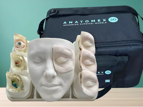
The Anatomex 3D Transorbital Surgical Simulation Model is a life-size, high-fidelity 3D training tool designed for surgeon education, skills acquisition, and patient explanation of transorbital approaches to the orbit and skull base.
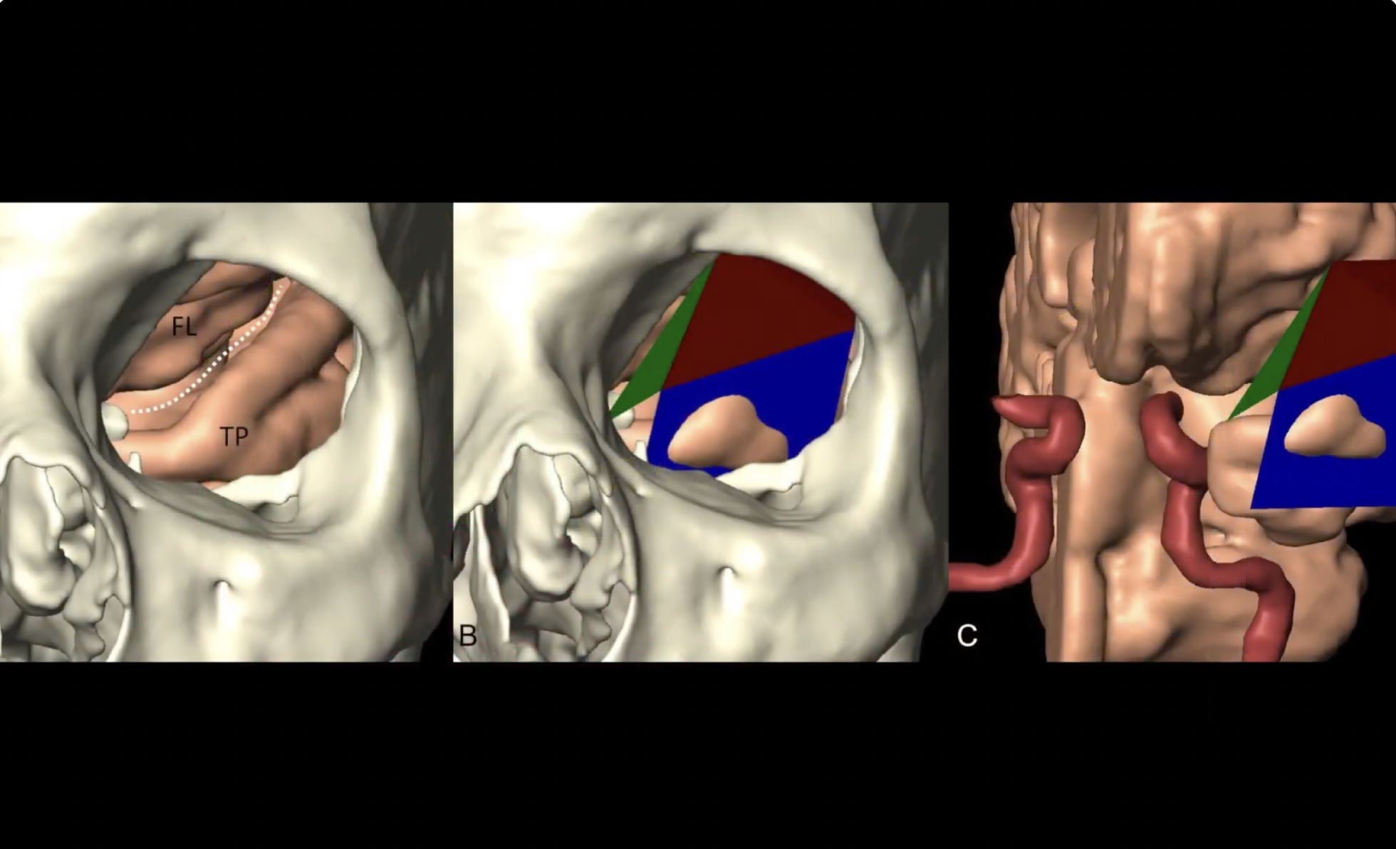
Developed in close collaboration with leading transorbital surgeons, the model accurately reproduces key bony and neurovascular anatomy, including:
- The bony orbit, skull base foramina, fissures, and optic canal
- The greater and lesser wings of the sphenoid, and the anterior clinoid process
- Access corridors to the middle cranial fossa, cavernous sinus, Meckel’s cave, and temporal lobe
- Intraorbital relationships of cranial nerves III, IV, V₁–V₃, and VI
This allows surgeons to visualise and rehearse transorbital procedures using a dry-lab model, enabling consistent practice without cadaveric constraints and in any clinical or educational setting.
Key features
1) Modular orbit design
Each kit includes three teaching orbits and four surgical simulation orbits, allowing progressive learning from orientation to full procedural rehearsal:
Teaching orbits
- Transparent normal orbit – for orientation and teaching of normal orbital and sphenoid anatomy.
- Transparent lateral sphenoid recess (“Sternberg”) orbit – derived from DICOM data of a patient with a well-pneumatised sphenoid sinus; ideal for demonstrating access and repair of spontaneous CSF leaks from the lateral recess.
- Normal bony orbit with integrated soft tissues – includes rectus muscles and cranial nerves III, IV, V₁–V₃, VI for intraorbital anatomy teaching.
Surgical simulation orbits (x4)
- Four full-procedure orbits with integrated dura, cavernous sinus, and cranial nerves for hands-on practice of the superolateral transorbital route:
- 2 “Sternberg” canal variants
- 2 normal sphenoid variants
2) Realistic cranium and soft-tissue overlay
- Life-size cranium with rotatable mount and base plate for precise head positioning during training.
- Removable soft-tissue (“flesh”) overlay to simulate periorbital soft tissues and skin, improving realism for incision planning, corridor formation, and instrument navigation.
3) Lifelike surgical feedback
- Materials engineered to replicate bone drilling resistance and realistic soft-tissue handling.
- Clear definition of critical landmarks to support structured dissection and shorten the learning curve.
4) Practical for courses and individual practice
- Robust, reusable base cranium and mount designed for repeated use.
- Replaceable surgical simulation orbits supplied in packs (cost-effective per session).
- Suitable for hospital skills labs, simulation centres, private practices, and international hands-on courses.
Full Transorbital Surgical Simulation Model Kit
Includes:
- Cranium with removable soft-tissue overlay and rotatable mount
- 2 transparent orbits (normal sphenoid sinus + 'Sternberg' variant)
- 1 bony orbit with extraocular muscles, cranial nerves, and vessels for educational purposes
- 4 solid surgical simulation orbits with integrated soft tissues (2 Sternberg + 2 normal)
- A4 base plate with rim
- Headband for navigation sensor mounting
- Padded transorbital carry bag
Article Link
Abstract
Article Link
Abstract
Objective: The lateral transorbital approach (LTOA) is a relatively new minimal access skull base approach suited for addressing paramedian pathology of the anterior and middle fossa. The authors define target zones for this approach and describe a series of cases with detailed measurements of visual outcomes, including those obtained with exophthalmometry.
Methods: The authors performed a retrospective analysis of a consecutive series of LTOA patients. Seven target zones were identified: 1) the orbit, 2) the lesser sphenoid wing and anterior clinoid, 3) the middle fossa, 4) the lateral wall of the cavernous sinus and Meckel's cave, 5) the infratemporal fossa, 6) the petrous apex, and 7) the anterior fossa. The authors used volumetric analyses of preoperative and postoperative MR and CT imaging data to calculate the volume of bone and tumor removed and to provide detailed ophthalmological, neurological, and cosmetic outcomes.
Results: Of the 20 patients in this cohort, pathology was in zone 2 (n = 10), zone 4 (n = 6), zone 3 (n = 2), zone 1 (n = 1), and zone 5 (n = 1). Pathology was meningioma (n = 10), schwannoma (n = 2), metastasis (n = 2), epidermoid (n = 1), dermoid (n = 1), encephalocele (n = 1), adenoma (n = 1), glioblastoma (n = 1), and inflammatory lesion (n = 1). The goal was gross-total resection (GTR) in 9 patients, all of whom achieved GTR. Subtotal resection (STR) was the goal in 8 patients (5 spheno-orbital meningiomas, 1 giant cavernous sinus/Meckel's cave schwannoma, 1 cavernous sinus prolactinoma, and 1 cavernous sinus dermoid), 7 of whom achieved STR and 1 of whom achieved GTR. The goal was biopsy in 2 patient and repair of encephalocele in 1. Visual acuity was stable or improved in 18 patients and worse in 2. Transient early postoperative diplopia, ptosis, eyelid swelling, and peri-orbital numbness were common. All 9 patients with preoperative diplopia improved at their last follow-up. Seven of 8 patients with preoperative exophthalmos improved after surgery (average correction of 64%). There were no cases of clinically significant (> 2 mm) postoperative enophthalmos. The most frequent postoperative complaint was peri-orbital numbness (40%). There was 1 CSF leak. Most patients were satisfied with their ocular (84%-100% of patients provided positive satisfaction-related responses) and cosmetic (75%-100%) outcomes.
Conclusions: The LTOA is a safe minimal access approach to a variety of paramedian anterior skull base pathologies in several locations. Early follow-up revealed excellent resolution of exophthalmos with little risk of clinically significant enophthalmos. Transient diplopia, ptosis, and peri-orbital numbness were common but improved. Careful case selection is critical to ensure good outcome.
Article Link
Abstract
Objective: The endoscopic transorbital approach (ETOA) has been demonstrated to be a feasible ventral route to the petrous apex. Yet, it has been pointed to as a deep and narrow corridor for anterior petrosectomy; particularly, medialization of the instruments can become an issue when targeting the petroclival area. To overcome this limitation, an ETOA with orbital rim removal (ETOA-OR) has been suggested, but not de facto compared, with a transorbital approach without removal of the rim. This addition could augment the surgical exposure and freedom of movement when accessing the petrous apex area.
Methods: Five human cadaveric heads (10 sides) were dissected. First, anterior petrosectomy was performed via a conventional ETOA (without orbital rim removal). Second, en bloc removal of the orbital rim was performed, with enlargement of the orbital craniectomy and, subsequently, further drilling of the medial petrous apex. Qualitative and quantitative comparisons are provided. An illustrative surgical case is also shown.
Results: The transorbital route allowed the authors to perform an anterior petrosectomy in all specimens. The landmarks of bone removal are superposed onto those in the transcranial route. The ETOA-OR increased the volume of craniectomy (from 4.0 mL to 5.5 mL), the lateromedial angulation, and superoinferior angulation of the instruments within the petrous area. Thus, this approach improved the exposure of the medial petroclival area, allowing for an augmented petrosectomy (from 1.4 mL to 2.0 mL, 39.5% increase) and for increased maneuverability, both in the petrous area (from 44.1 cm2 to 76.5 cm2, 73.3% increase) and in the posterior fossa (from 20.2 cm2 to 52.0 cm2, 158% increase). The ETOA-OR was also pragmatically applied to treat a recurrent petroclival meningioma. Complete removal was achieved, the abducens nerve palsy improved, and the trigeminal neuralgia decreased in severity, yet still required medication.
Conclusions: The authors provide the first formal anatomical comparison between the transorbital approach with preservation of the orbital rim and a transorbital approach with removal of the rim to access the petrous apex. In addition, an illustrative case is used as a proof of concept and feasibility. According to the authors' data, the ETOA-OR significantly improves surgical exposure and the surgeon's comfort in this deep region. The bony defect can be reconstructed to avoid cosmetic deformities, maintaining the minimally disruptive concept of transorbital surgery.
Article Link
Abstract
Background: The endoscopic superior eyelid approach is a relatively novel mini-invasive technique that is currently investigating for skull base cancers. However, questions remain regarding specific approach-related complications when treating different skull base tumors. This study aims to analyze any surgical complications that occurred in our preliminary consecutive experience, with specific focus on orbital outcome.
Methods: A retrospective and consecutive cohort of patients treated via a superior eyelid endoscopic transorbital approach at the Division of Neurosurgery of the Hospital Clinic in Barcelona was analyzed. Patients features were described in detail. Complications were divided into 2 groups to analyze separately the approach-related complications, and those resulting from tumor removal. The ocular complications were subdivided into early ocular status (<3 weeks), late ocular status (3-8 weeks), and persistent ocular complications. The "Park questionnaire" was used to determine patient's satisfaction related to the transorbital approach.
Results: A total of 20 patients (5 spheno-orbital meningiomas, 1 intradiploic Meningioma, 2 intraconal lesions, 1 temporal pole lesion, 2 trigeminal schwannoma, 3 cavernous sinus lesions, and 6 petroclival lesions) were included in the period 2017-2022. Regarding early ocular status, upper eyelid edema was detected in all cases (100%) associated with diplopia in the lateral gaze in 30% of cases, and periorbital edema in 15% of cases. These aspects tend to resolve at late ocular follow-up (3-8 weeks) in most cases. Regarding persistent ocular complications, in one case of intraconal lesion, a limitation of eye abduction was detected (5%). In another patient with intraconal lesion, an ocular neuropathic pain was reported (5%). In 2 cases of petroclival menigioma, who were also treated with a ventriculo-peritoneal shunt, slight enophthalmus was observed as a persistent complication (10%). According to the Park questionnaire, no cosmetic complaints, no head pain, no palpable cranial irregularities, and no limited mouth opening were reported, and an average of 89% of general satisfaction was encountered.
Conclusions: The superior eyelid endoscopic transorbital approach is a safe and satisfactory technique for a diversity of skull base tumors. At late follow-up, upper eyelid edema, diplopia, and periorbital edema tend to resolve. Persistent ocular complications are more frequent after treating intraconal lesions. Enophthalmus may occur in patients with associated ventriculo-peritoneal shunt. According to patient's satisfaction, fairly acceptable results are attained.
A way to improve skull base surgery through the advanced application of endoscopic techniques.