High-fidelity teaching and skills training for endoscopic transorbital approaches
The Anatomex 3D Transorbital Surgical Simulation Model is a life-size, high-fidelity 3D training tool designed for surgeon education, skills acquisition, and patient explanation of transorbital approaches to the orbit and skull base.
Developed in close collaboration with leading transorbital surgeons, the model accurately reproduces key bony and neurovascular anatomy, including:
- The bony orbit, skull base foramina, fissures, and optic canal
- The greater and lesser wings of the sphenoid, and the anterior clinoid process
- Access corridors to the middle cranial fossa, cavernous sinus, Meckel’s cave, and temporal lobe
- Intraorbital relationships of cranial nerves III, IV, V₁–V₃, and VI
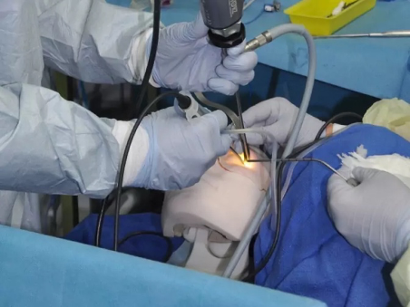
This allows surgeons to visualise and rehearse transorbital procedures using a dry-lab model, enabling consistent practice without cadaveric constraints and in any clinical or educational setting.
Key features
1) Modular orbit design
Each kit includes three teaching orbits and four surgical simulation orbits, allowing progressive learning from orientation to full procedural rehearsal:
Teaching orbits
- Transparent normal orbit – for orientation and teaching of normal orbital and sphenoid anatomy.
- Transparent lateral sphenoid recess (“Sternberg”) orbit – derived from DICOM data of a patient with a well-pneumatised sphenoid sinus; ideal for demonstrating access and repair of spontaneous CSF leaks from the lateral recess.
- Normal bony orbit with integrated soft tissues – includes rectus muscles and cranial nerves III, IV, V₁–V₃, VI for intraorbital anatomy teaching.
Surgical simulation orbits (x4)
- Four full-procedure orbits with integrated dura, cavernous sinus, and cranial nerves for hands-on practice of the superolateral transorbital route:
- 2 “Sternberg” canal variants
- 2 normal sphenoid variants
2) Realistic cranium and soft-tissue overlay
- Life-size cranium with rotatable mount and base plate for precise head positioning during training.
- Removable soft-tissue (“flesh”) overlay to simulate periorbital soft tissues and skin, improving realism for incision planning, corridor formation, and instrument navigation.
3) Lifelike surgical feedback
- Materials engineered to replicate bone drilling resistance and realistic soft-tissue handling.
- Clear definition of critical landmarks to support structured dissection and shorten the learning curve.
4) Practical for courses and individual practice
- Robust, reusable base cranium and mount designed for repeated use.
- Replaceable surgical simulation orbits supplied in packs (cost-effective per session).
- Suitable for hospital skills labs, simulation centres, private practices, and international hands-on courses.
Full Transorbital Surgical Simulation Model Kit
Includes:
- Cranium with removable soft-tissue overlay and rotatable mount
- 2 transparent orbits (normal sphenoid sinus + 'Sternberg' variant)
- 1 bony orbit with extraocular muscles, cranial nerves, and vessels for educational purposes
- 4 solid surgical simulation orbits with integrated soft tissues (2 Sternberg + 2 normal)
- A4 base plate with rim
- Headband for navigation sensor mounting
- Padded transorbital carry bag